Have you ever wondered what your brain looks like? I mean, what really is that mush between your ears anyways? I think it's natural to wonder about this organ that controls every single thing you do, even the automatic things, but as a Brain & Cognitive Sciences (BCS) major, for me it's more than just wonder—it's unadulterated fascination.
Fortunately for me, I go to a stellar research university that is more than well-equipped to give me a concrete answer to my fascination.
Anyone who takes a BCS class here will receive a form on the first day of lecture asking if they want to be put on an email list that will notify them of studies in which they can participate. I've taken so many BCS classes at this point that I get a seemingly never-ending stream of emails from the department. Most of them I just don't even look at anymore because invariably they're about language studies, which are insanely boring to participate in. But a few weeks ago, I got an email about an fMRI study that caught my eye.
"The task is very simple," it said. "You would be lying in the scanner and actively paying attention to images of tools, animals, famous places, and famous faces [. . .] The whole thing takes about an hour and 15 minutes. You will be compensated $20 for completing this."
Having an opportunity to see my brain and make $20 at the same time sounded like the ideal opportunity. In reality, they probably could've gotten me to pay them $20 just to see my brain . . . but I'm glad it was the other way around.
I'd never had an MRI or fMRI before so I was a little bit nervous prior, but mostly I was just excited. I know this is really odd, but I ultimately found the whole experience really relaxing—lying down on a comfy bed in a tiny space. And I didn't even really have to do anything, just look at pictures of things like the Eiffel Tower, Alec Baldwin, and a wrench.
The grad student who was running the test was really receptive to my interest in the study, and later that day sent me digital images of my brain—both in the neutral and testing states.
This is my brain in the neutral state—check this insanity out:

THAT'S MY ACTUAL BRAIN. How crazy is that? I would also just like to take this moment to congratulate myself on having a stunning corpus collosum.
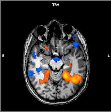
This is my brain during the testing state:

The orange areas represent regions of the brain that are implicated in recognizing animals, whereas the blue regions are implicated in tool recognition.
I've been looking at images like this since my senior year of high school when I took my first neurobiology course, but the images have always been in textbooks and are of an unnamed person's brain. Part of the reason why I found this experience so exciting is because this image represents a fact about brain differentiation that I've learned over and over again in my classes—that tools and animals have distinct processing areas in the brain—but here I got to see that very principle in the context of my brain.
That's some pretty valuable experiential learning right there. It's basically like saying, if you don't want to take your textbook's word for something, or even your professor's or TA's, just head over to the Rochester Center for Brain Imaging (RCBI) and see it in action in your own brain. And it's pretty cool that I was able to do that on my own campus; it didn't even take me 5 minutes to walk to the RCBI, where I actually ran into two undergraduate friends of mine who do research there.
On that note, I'm gonna go study for my Neurobiology final. . . .